Tendonitis Surgical Photos
Before and After Release of the Retinaculum
![A surgical opening where the foreskin of a knee is peeled back revealing internal flesh](https://lincoln-ne.carpaltunnelrelief.net/wp-content/uploads/sites/10/2018/07/De_Quervains_1.jpg)
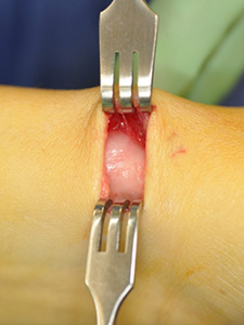
Before Tendonitis Relief Surgery
The thick extensor retinaculum (sheath) that needs released as it pinches tendons.
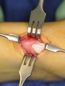
![A surgical opening where the foreskin of a knee is peeled back revealing internal flesh](https://lincoln-ne.carpaltunnelrelief.net/wp-content/uploads/sites/10/2018/07/De_Quervains_2.jpg)
During Surgery
A partial release of the retinaculum.
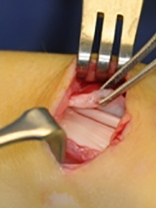
![A surgical opening where the foreskin of a knee is peeled back revealing internal flesh](https://lincoln-ne.carpaltunnelrelief.net/wp-content/uploads/sites/10/2018/07/De_Quervains_4.jpg)
After Surgery
Complete release of the retinaculum. Note how thick this abnormal band is. It should be thin like cellophane.